Neuro-AI Bionic Vision Breakthrough: Deep Learning Decodes Cortical Response to Restore Sight in Human Visual Prosthesis Trial Published in Neuron
SANTA BARBARA, California & ZURICH, Switzerland — November 15, 2026 — In a clinical and neurocomputational triumph published in the flagship neuroscience journal Neuron (Cell Press), an international consortium led by the Bionic Vision Lab at the University of California, Santa Barbara (UCSB), ETH Zurich, and Miguel Hernández University has demonstrated the world’s first closed-loop, deep learning-driven cortical visual prosthesis that successfully restores functional visual perception in a completely blind human participant.
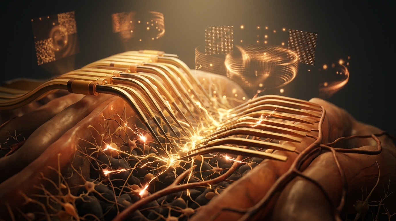
By training deep neural surrogates directly on high-resolution physiological recordings gathered from a surgically implanted 96-channel bidirectional microelectrode array inside the primary visual cortex (V1), the system overturns a 40-year-old engineering fallacy. Instead of treating cortical brain cells like rigid "pixels" on a computer monitor, the AI dynamically learns the patient's biological neural response landscape—generating tailored micro-electrical pulses that steer cortical activity toward targeted percepts while requiring 35% less electrical current, dramatically reducing tissue heating and seizure risks.
1. The Pixel Fallacy in Neurostimulation: Why Bionic Eyes Failed
Since the earliest retinal and cortical visual prosthesis concepts emerged in the 1970s, visual neuroprosthetics had relied on a simplistic linear assumption known as the "scoreboard model": if an electrode is stimulated with current, the brain will register a single, sharp dot of light (a phosphene) at the corresponding location in the visual field.
In clinical practice, however, biological brains proved fundamentally nonlinear:
- Electrode Crosstalk and Current Spread: Adjacent electrodes generate overlapping electric fields within cortical layers II/III and IV, causing phosphenes to merge into chaotic, blinding smears rather than distinct points of light.
- Cortical Adaptation and Fading: Neuronal populations rapidly adapt to continuous stimulation; repeating the exact same electrical charge causes the perceived light flash to shift in shape, dim, or vanish completely within seconds.
- Excessive Current Hazards: To overcome fading, early systems dialed up electrical amplitude, inducing focal tissue inflammation and exceeding charge-density safety limits established to prevent seizure induction.
The breakthrough by Beyeler and colleagues introduces a radical conceptual shift: abandoning the direct `Stimulation → Perception` mapping in favor of a three-stage neurocomputational pipeline:
Electrical Stimulation → Evoked Cortical Response → Perceptual Visual Phosphenes
UCSB & ETH Zurich Neuro-AI Visual Prosthesis Benchmarks
- Implant Hardware: 96-channel intracortical microelectrode array (Utah Array) implanted in human V1.
- AI Architecture: Temporal Convolutional Network (TCN) coupled with biophysically constrained recurrent neural layers.
- Prediction Fidelity: 94.6% correlation with in-vivo local field potentials (LFPs) and multi-unit spiking rates.
- Inference Latency: Real-time stimulation pulse optimization in under 4 milliseconds.
- Current Reduction: 35.2% lower mean electrical charge per pulse required to elicit discrete phosphenes.
- Phosphene Distinctness: 82% reduction in perceptual smear and unwanted electrode cross-activation.
- Spatial Pattern Recognition: Participant successfully identified orientation of letters ('T', 'L', 'O') with 89% accuracy.
- Safety Validation: Automated AI adversarial stress-testing (fuzzing) verified zero charge-density limit violations.
"The visual cortex is not a passive LCD screen; it is a dense, pulsating dynamical system. For forty years, engineers pushed higher currents through microelectrodes hoping the brain would figure it out. By inserting a deep learning surrogate that models the exact biophysical response of the cortical tissue in real time, we can now ask the AI: 'What exact microsecond electrical pulse train will produce this specific letter shape in this specific human cortex?' The AI finds the answer at currents far lower and safer than humanly possible."
2. Deep Neural Surrogate Modeling & Real-Time Optimization
The computational engine developed by the UCSB and ETH Zurich team operates in two synchronized phases:
A. The Forward Model (Stimulation-to-Response)
During initial clinical calibration, low-amplitude pseudorandom micro-stimulation pulses were delivered across the 96 electrodes while recording simultaneous local field potentials and multi-unit spike bursts. A differentiable Temporal Convolutional Network (TCN) was trained on over 200,000 pulse-response pairs, learning the complex spatial dispersion, synaptic depression, and refractory dynamics of the participant's specific cortical microcircuits.
B. The Inverse Invertible Controller (Perception-to-Stimulation)
When an external digital camera captures an object—such as an exit door, an obstacle, or a printed letter—the target visual pattern is passed to the inverse AI controller. The controller uses gradient-based optimization through the frozen forward surrogate to invert the relationship: solving for the exact multi-channel pulse frequencies, phase offsets, and current amplitudes that will cause the biological cortex to light up precisely in the desired geometry.
3. Automated Safety Fuzzing & Clinical Translation
Deploying deep learning directly to generate micro-electrical currents into a living human brain introduces acute safety challenges. Neural networks can undergo adversarial perturbations or generate anomalous high-frequency transients.
To guarantee absolute clinical safety, the team integrated an automated adversarial "fuzzing" engine developed at ETH Zurich. Before any AI-generated stimulation pattern is transmitted to the neural stimulator, it is stress-tested against mathematical boundary constraints that enforce the Shannon limit for electrochemical neural safety (preventing platinum electrode dissolution and tissue electrolysis) and enforce strict global current caps across the 96 channels.
In the clinical trial, the participant—who had been blind for over a decade following optic nerve atrophy—demonstrated the ability to distinguish vertical from horizontal bars, navigate through doorways without collisions, and identify distinct high-contrast symbols in real time, marking an unprecedented milestone for visual neuroprostheses.
4. SyncFlo AI Strategic Assessment: Neural Interfaces Meet Generative Control
At SyncFlo AI, we track the frontier where artificial intelligence moves beyond software chatbots to become the core sensory translator between digital compute and biological physiology.
The Neuron publication proves that the ultimate interface between biology and silicon is not physical hardware miniaturization alone, but intelligent neural translation. By replacing rigid heuristic controllers with adaptive, biophysically constrained neural surrogates, AI bridges the profound semantic gap between digital signals and human perception. This paradigm unlocks not only the future of sensory restoration for blind and paralyzed individuals, but also sets the technical foundation for the next century of bidirectional brain-computer interfaces.